
Dr. John Reinisch rebuilds the outer ear. Dr. Ronen Nazarian restores the hearing behind it. Together, they plan your child’s care from the first scan to the final follow-up, and often complete both in a single outpatient surgery.
Most children with microtia also have a closed ear canal. Building the outer ear is plastic surgery. Opening the canal and rebuilding the eardrum and middle ear is ear surgery. Instead of treating these one after the other in separate offices, we plan both from day one.
What microtia is, and what it isn’t.
Microtia means “small ear.” A child is born with an outer ear that is undersized, incompletely formed or absent. It usually affects one side, most often the right. About one child in five has it on both sides.
It is uncommon, appearing in about 1 in 8,000 to 10,000 births in the United States, and is seen more often in boys. Routine prenatal scans rarely examine the outer ear, so most parents first learn about it in the delivery room.
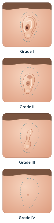
The grade helps guide the plan, but every ear is assessed individually.
The ear is smaller than usual, but its main folds are recognizable. The canal may be narrow or present.
Parts of the ear, usually the upper portion, are missing or underformed. The canal is often narrow or closed.
The most common form: a small, peanut-shaped remnant of skin and cartilage, typically with no ear canal.
No outer ear and no canal.
Children with one hearing ear often seem to manage well, so the loss is easy to overlook. It shows up later, in noisy classrooms, group conversations and on the playground.
Common signs include:
Most children with microtia also have aural atresia, meaning the ear canal is closed or very narrow. The eardrum and the tiny middle ear bones may also be underdeveloped. Sound cannot travel through, which causes conductive hearing loss on that side.
The inner ear, which turns sound into nerve signals, usually works normally. That is good news, because hearing can often be restored by opening a path for sound.
There is no single right answer for every child. We walk you through each approach, including the ones we perform less often, and help you weigh them against your child’s anatomy, age, and hearing needs.
A custom-shaped porous polyethylene framework is placed where the ear should be. It is covered with a thin, well-vascularized layer of the child’s own tissue from the scalp, then with a skin graft. Blood vessels grow into the framework’s pores and anchor it as part of the body.
Usually completed in one outpatient surgery
Can be done from about age 3, before a child starts school
No rib cartilage removed and no chest scar
Ear shape and projection are designed to match the other side
Reconstruction plus hearing, one session.
For children whose CT shows suitable anatomy, Dr. Nazarian creates a new ear canal and eardrum and reconstructs the middle ear, while Dr. Reinisch builds the outer ear. The new canal also helps position the reconstructed ear naturally.
A custom silicone ear, made by an anaplastologist, is attached with adhesive or small implanted magnets. It can look very realistic, but it must be removed and cleaned daily, replaced every few years, and it does not improve hearing. We are glad to refer families who prefer this route.
If the anatomy does not favor canal surgery, sound can be sent to the inner ear through the bone of the skull. Options range from soft headband devices for infants to implanted bone-conduction systems. Where possible, the implant is placed through the same incision used for the ear reconstruction, so no new scar is added.
Cartilage is taken from the child’s ribs and carved into an ear framework. It is a well-established method, and we discuss it openly. Families should know what it usually involves:
The right next step depends on where your child is today.
| Your child is? | What usually happens now? | What to plan for? |
| A newborn or infant | Hearing tests on both sides; a soft-band hearing device if hearing is reduced | An early consultation to map out the years ahead |
| A toddler (1–3) | Speech and hearing monitoring; temporal bone CT at around 2½ | Deciding between canal surgery and a hearing device |
| Preschool age (3–5) | Ear reconstruction, often combined with hearing surgery | Finishing treatment before kindergarten |
| School age or a teen | Reconstruction and hearing options are all still open | Fitting surgery around school breaks; involving your child in the decision |
| An adult | Same options as children; many adults choose a single combined surgery | Time off work, usually 1–2 weeks |
| Already treated elsewhere | Review of previous surgery, photos and scans | Revision or a new reconstruction, depending on the tissue available |
Microtia treatment is designed to restore both the appearance of the outer ear and, when possible, functional hearing.
Many children with microtia also have aural atresia, a closed or narrow ear canal. Rebuilding the ear and the canal as part of one plan can make a real difference to hearing. The sooner sound reaches the inner ear, the better a child is supported in learning to speak, understand language and keep up at school.
Microtia care has two goals: an outer ear that looks natural, and, wherever possible, hearing that works. Treating both early and with careful planning can help a child grow, communicate and feel confident in the years ahead.
With implant-based techniques such as MEDPOR®, Dr. Reinisch shapes a new ear to mirror the other side in size, outline and how far it stands out from the head. When the face looks balanced, children often feel more at ease with friends and classmates and less aware of being different.
When treatment happens at the right age, children can start school with an ear that looks natural and hearing that works better. Parents often tell us that acting early brings peace of mind and lets their child take part fully in everyday life.
Recovery from ear reconstruction is relatively short but needs to be handled carefully. We prescribe medications to minimize discomfort and prevent infection, as well as provide all resources necessary for a full recovery.
| When | What happens |
| Surgery day | Home the same day with a protective dressing. Rest, simple pain relief, no sleeping on the treated side. |
| Days 2–3 | First check. The outer dressing comes off and swelling starts to settle. |
| Week 1 | Most children are back to quiet routines and school with the surgeon’s approval. |
| Weeks 2–4 | Splint and stitches removed in stages. Parents learn gentle cleaning and how to use the night-time ear mold. |
| Weeks 4–6 | Return to sports and active play, with normal protective gear. |
| Month 3 onward | Swelling has largely resolved and the final shape is visible. Hearing is reviewed with doctor. |