
Hearing that your child was born without an open ear canal raises urgent questions. Will they hear? Will they talk on time? Is something else going on? This page explains what aural atresia is, how it’s found and measured, and what to watch for as your child grows.
For an overview of all microtia and atresia treatment, see Microtia & Aural Atresia Care.
The ear canal is the passage that carries sound from the outer ear to the eardrum. In aural atresia, that passage didn’t form, or formed only partly. It’s one of a family of related conditions:
Because sound can’t pass through, the result is conductive hearing loss on that side. Behind the closed canal, the eardrum and middle ear bones may be small, joined together or missing. The inner ear usually formed normally.
The outer ear, ear canal and middle ear all form early in pregnancy, from the same small cluster of tissue that also shapes the jaw and parts of the face. For a while, the developing canal is filled with a plug of cells. That plug normally clears in the later months of pregnancy and leaves an open passage. When this process is interrupted, the canal stays closed or narrow.
This is why atresia so often appears together with microtia, and sometimes with differences in the jaw or face. In most children no single cause is found, and it isn’t linked to anything parents did or didn’t do.
Most children have atresia on its own or with microtia only. In some, it’s part of a wider pattern, such as:
When other features are present, a genetics consultation can clarify the diagnosis and what it means for future children. Some of these conditions also affect how well canal surgery is likely to work, which is why we look at the whole child, not just the ear.
| At birth | No visible ear canal opening, or a very small one An outer ear that is small or differently shaped A newborn hearing screen that doesn’t pass on one or both sides |
| As your child learns to talk | Children with hearing in one ear usually develop speech normally, but keep an eye on these milestones and mention any concerns at your next visit: By 12 months: not yet pointing, waving or using other gestures Children with atresia in both ears need hearing support from the first weeks of life, so speech can develop on time. |
| If the canal is narrow rather than closed | A narrow canal can trap skin and wax behind it. Over time this can form a cholesteatoma, a growth of trapped skin that slowly damages surrounding bone. Call us if your child has:
Children with stenosis need regular checks even if they hear well. |
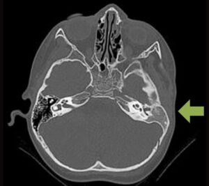
Hearing is checked in stages, from the first days of life to a CT scan at around age 2½.”
| When | What happens |
| First days to first months | Newborn hearing screening, then an auditory brainstem response (ABR) test. This measures hearing in each ear while the baby sleeps. |
| From about 6 months | Behavioral hearing tests: an audiologist watches how your child responds to sounds and speech. |
| Around age 2½ | A temporal bone CT scan maps the canal, middle ear, and inner ear. It’s done earlier if a narrow canal raises concern about cholesteatoma. |
| Any time, if needed | Genetic evaluation when other facial or body features are present. |
Surgeons often use the Jahrsdorfer scale to judge whether a new canal is likely to give useful hearing. Each structure that’s present and well formed earns points:
What the score means: generally, 7 or above suggests a good chance of strong hearing after canal surgery, 6 is borderline, and 5 or below usually points toward a hearing device instead. The score guides the decision but doesn’t make it on its own.
| Structure on CT | Points |
| Stapes (the smallest hearing bone) | 2 |
| Oval window open | 1 |
| Middle ear space present | 1 |
| Facial nerve in a normal position | 1 |
| Malleus and incus present | 1 |
| Mastoid air cells developed | 1 |
| Incus joined to the stapes | 1 |
| Round window present | 1 |
| Outer ear appearance | 1 |
| Total possible | 10 |
Most children with atresia grow up to hear, speak and learn well, especially when hearing is checked early and supported where needed. Hearing in the affected ear may not become completely normal, but treatment often brings it close. Regular follow-up makes sure any change is caught early, especially in children with narrow canals.
Our FAQ covers diagnosis, hearing, surgery, recovery, and travel.
Yes. Some children have a closed or narrow canal with a normal-looking outer ear. They still need the same hearing tests and imaging.
Not necessarily. A narrow canal may let some sound through, but it can trap skin and lead to cholesteatoma, so it needs regular monitoring.
Usually not. Balance is controlled by the inner ear, which typically forms normally in atresia.
No. A closed canal doesn’t cause pain on its own. New pain, discharge or odor in a narrow canal should be checked promptly.
Yes. Hearing evaluation, canal surgery and hearing devices are all options at any age.
Once the CT shows what’s behind the closed canal, there’s almost always a way to bring sound through. Dr. Nazarian and Dr. Reinisch will help you choose the path that fits your child’s anatomy, age and daily life. Every option is explained at your consultation, including what to expect for hearing, recovery and long-term care.